The Course of Time: Varicose Veins Through the Ages

Evolution is a wonderful thing, but varicose veins are one associated aspect that humans could have happily lived without. Unfortunately, the ability to stand and walk on two legs came with unsightly and unhealthy consequences. But if you think vanity associated with the current “selfie age” triggered a deeper dislike of such visual flaws, think again. Humans have been self-conscious about their varicose veins for thousands of years. And as far as we know, treatments have been around — some more rudimentary than others — since 1550 BCE. We’re fortunate these days to have access to non-invasive medical techniques, but imagine for a moment, living centuries ago with varicose veins — what would you have done, and how would you have been treated? If you think you’re unlucky to have varicose veins, read on to understand the history of varicose veins and find out what people went through before the dawn of modern medical procedures. You might just see your plight in a different light.


Chances are, you stumbled upon this blog while researching your own venous situation. Of course, when you have a condition, it pays to know as much about it as you can, for your own peace of mind. So for a moment, let’s briefly revisit the basics: What are varicose veins? They are:

  • Abnormally large, tortuous veins that typically appear in the legs;
  • A cause of aching, swelling, itching, leg heaviness, and skin ulceration
 

So how far have humans come in the research and treatment of varicose veins? And what did people face when they needed treatment? The earliest record of varicose veins is in the papyrus of Ebers, written 1550 BCE, where the author described them as “tortuous and solid, with many knots, as if blown up by air”, and recommended people leave their veins in place.

Ebers Papyrus
Ebers Papyrus – 1500 BC
 

The first illustration of a varicose vein dated from 400 BCE, a votive tablet found at the base of the Acropolis — so that gives you an extra idea just how long varicose veins have been bothering people. It was around this time that Hippocrates, the “Father of Medicine”, suggested people do something about their varicose veins, because he noticed a correlation between veins and leg ulcers. This was the introduction of vein punctures, cautery (using a hot or caustic agent) and compression bandages as a treatment.

ancient depiction of varicose veins from Acropolis
Depiction of a varicose vein – 400 BC
 

Over the next few centuries, the developments in treatments were many and varied. For example, in 270 BCE, two Egyptian physicians made surgery possible through the invention of forceps to block veins and control bleeding. But a low point in surgical history was around 0 CE, when the removal of a varicose vein was too much for even a notorious Roman warlord. He had one leg done and opted out of having the other fixed, saying “I see the cure is not worth the pain.” Today easier, more advanced varicose vein treatments are available with no down time.


Mercifully, surgical developments in varicose vein treatment continued. A Greek surgeon realized around 600 CE that the great saphenous vein — the longest vein in the human body — could be ligated or removed. Then in 1485, a landmark development was when Leonardo da Vinci produced accurate drawings of lower-limb veins, helping medical world make sense of how the venous system worked.
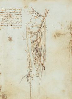
da Vinci's anatomical drawing
Leonardo da Vinci – 1485 AD
 

Two centuries after da Vinci’s drawings were rendered, the first documented attempt at sclerotherapy took place — acid is injected to create thrombus — a blood clot. This set the scene for the 1800s, where a flurry of medical developments were made. Charles Gabriel Pravaz invented his injection syringe made of glass, rubber and leather, and Francis Rynd followed up with the hypodermic needle.


By the 1900s, critical tools of the trade had been invented, and surgical processes were now being refined and published. Options included saphenous vein ligation; vein perforation to treat ulcers; and application of a special agent to close varicose veins. The links between chronic pelvic pain and vein congestion in that region were discovered, known as pelvic venous insuficiency, and Sven-Ivar Seldinger figured out a technique to access veins using guidewires.


Enter the modern age of venous treatment. Since the 1960s, treatment of vascular disease has included the use of guidewires, catheters, angioplasty and stents. If you had vascular issues these days, you were fortunate to have surgeons with many cutting-edge treatment options at their disposal. More recent and sophisticated treatments tended to have impressive scientific titles, such as duplex ultrasound scanning, radiofrequency ablation, foam sclerotherapy, and diode laser.
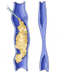
sclerotherapy
Sclerotherapy uses an injectable medication to close varicose veins.
 

Which brings us to now. Do you have a vascular disorder that concerns you? Rest assured, medical development has never stopped. Perhaps your treatment might include a ClariVein device, which can be used without tumescent anesthesia. Asclera is an option for closing small veins. And VenaSeal is a system that closes veins using an adhesive agent. Your specialists know just the right treatment for you and you have nothing to worry about. The days of the painful surgery that so terrified the Roman warlord are thankfully a thing of the very distant past.

  history of varicose veins infographic

Share this Image On Your Site

What To Expect During Your Ultrasound

Some very common questions new patients have when they come in for their first appointment are “What are you looking for? How can you tell if I have varicose veins?”. Ultrasound is excellent for diagnosing varicose veins and is used throughout the treatment plan. Patients can expect to have an ultrasound at all of their appointments.

When the exam is started, the sonographer adjusts the bed to what’s referred as the reverse Trendelenburg position, which is with the foot of the bed much lower than the head of the bed. This simulates standing, which increases venous pressure on the legs, and the blood flow patterns can be evaluated more accurately than laying down flat on a bed, which creates 0% hydrostatic pressure and the flow patterns would be minimalized. The patient does stand for part of the ultrasound when the back of the legs are being evaluated.

The sonographer evaluates all the deep and superficial veins in the legs. The legs are checked for any superficial or deep blood clots. The starting and ending point of all the veins are evaluated, and the flow checked for reflux. Reflux in the veins is when blood is going in the wrong direction (towards the feet) when it should be flowing up towards the heart. The heart pumps the blood from the arteries to the

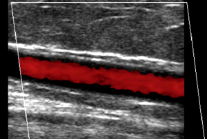
ultrasound image demonstrating venous reflux using color doppler
Reflux in a vein identified using color doppler.

extremities, and the veins take the blood back to the heart. The amount of blood returning to the heart varies at any moment, as this is achieved by breathing. When there is increased abdominal pressure, for instance, taking a deep breath, the diaphragm presses down on the lower venous system, decreasing flow, just for a moment, and when breath is exhaled, the pressure is released from the lower venous system, and the blood rushes back to the heart. This happens over and over.

There are valves throughout the veins, and when there is increased pressure on the veins, the valves will stop the blood from going back to the feet. When you have chronic venous insufficiency syndrome, the valves have stopped working and every time there is increased abdominal pressure, the valves won’t hold the blood where it is, and it all rushes towards the feet. This phenomenon can be visualized on ultrasound by using doppler, which can tell what direction the blood is flowing. The sonographer has the patient reproduce this through the Valsalva movement (bearing down) multiple times throughout the procedure. This is how venous disease is diagnosed.

A week or so after the procedures another ultrasound is performed, this time to check for vein closure and to make sure there are no complications or blood clots. After several months the body has absorbed the vein and it is not visible on ultrasound anymore.

Ultrasound is a very useful modality in the diagnosis of varicose veins and has revolutionized the treatment and management of venous insufficiency syndrome.

Varicose Vein Treatment: Understanding What Your Insurance Will Cover

Dealing with treatment of varicose veins can be tricky when it comes to prior authorization with an insurance company. There are many guidelines that need to be followed and they are specific to each insurance company. Examples including vein size, types of veins, pain level, compression stockings and Nsaid use are just a few that each insurance will look for at the time a prior authorization is submitted.

Pre-Authorizing Treatment

At IVC we are very aware of the importance of obtaining the correct measurements and making the patient aware of their responsibility before an authorization can be obtained. No one wants to go into a procedure without being sure that the insurance will pay their part. We want our patients to be relaxed on their procedure date, not stressed about whether they will have an issue with their insurance later on. Because patient care is so important to us, we pride ourselves in knowing what the insurance company needs, we have an open relationship with many insurance companies who appreciate our clinical notes and ultrasound reports and have open communication with us to explain any changes because we are not trying to sneak past their requirements.

Knowing Your Out-of-Pocket Expense

Not only can you have the confidence in our authorization department, we offer what many clinics do not, knowing what your out of pocket cost is before starting the varicose vein treatments. Knowing what your portion of the treatment will cost you will help you decide if the treatment is possible, and prepare with your HSA or Flex Spending accounts. You will hopefully better understand what your deductible, co-insurance and out of pocket max are and how they apply to the treatments you receive at IVC.

Getting started with your new patient appointment will not only educate you on insurance requirements and cost but will also get you started with those requirements and be ready if your deductible is met for the year.

Varicose Vein Procedures: Endovenous Thermal Ablation

Endovenous Thermal Ablation

The first procedure typically performed is the venous ablation. This is where the vein is burned with either laser or radio frequency catheters. During this procedure, the patient is sedated with valium, nitrous oxide, or both. The patient can choose the level of sedation they prefer.

During this procedure, the sonographer will map the vein to be ablated and mark a location that appears to be a good access point. What we look for is whether the vein is straight and has a large diameter, which makes accessing the vein easy. Veins will sometimes spasm or be a smaller diameter early in the morning when the patient hasn’t been standing or active all day, or when the leg is cold. In these cases, we use gravity, a Nitroglycerin patch, and a blanket to help warm up the leg and increase the diameter of the vein.

Once a location is marked, we prep the leg with a cleansing/sterilizing solution and set up the procedure using sterile technique. We ask that the patient not touch the sterile field, so to prevent infection. When the physician enters the room, he will also dress in a sterile gown and will don sterile gloves. We take every precaution to keep the patient safe and infection free.

At this point, the physician will access the vein and start the procedure. Once the vein is accessed, a “guide” wire is inserted and the sonographer will follow the wire up the vein to make sure we are in the vein and not in the surrounding tissue. After that, the catheter, with the heating element, is inserted and once again the sonographer follows the catheter with ultrasound to make sure it is in the vein to be treated.

Once the catheter is an acceptable distance from the deep venous system, we need to numb around the vein. This is two-fold: The first reason is to numb and protect the tissue surrounding the treated vein from being burned. The second is to compress the vein around the catheter so the walls of the vein will be treated.

Once this all complete, the catheter is slowly removed and the vein will be ablated. We close the access point with steri-strips and the procedure is finished. The patient is then required to wear a compression stocking for one week.

Prevalence of Varicose Veins

If you suffer from varicose veins, you are not alone. Studies suggest that approximately 40 million people in the United States have variscosities. Half of this group are over the age of 50 and more than 60 percent are older women. Hormone changes throughout life tend to make women more susceptible to this condition. The problem may also develop due to genetics. Additionally, being overweight can also be a contributing factor. Anyone having an occupation that requires standing for extended periods or chronically lifts heavy weight may also be at higher risk.

Varicose Veins Causes and Prevention

While there is nothing that one can do about family history, and changing occupations may be difficult, other lifestyle changes may at least serve to delay varicose vein development. Maintaining a healthy weight and getting sufficient amounts of exercise weekly keep muscles toned and help leg veins perform more efficiently. Physicians at The National Institutes of Health also recommend that if needing to stand or sit for long lengths of time at a job, take breaks by changing positions. Avoid crossing your legs while sitting and elevate legs when sitting or just lounging around at home. Physicians at the Varicose Vein Center in Utah explain that the condition involves venous blood vessels that become engorged, swollen and sometimes twisted because valves within the veins falter. Veins return blood to the heart and lungs for oxygenation. The blood travels upward from the legs through the assistance of contracting leg muscles. Valves in the vessels keep the blood from flowing in reverse. When the valves weaken or completely fail, blood flow stalls and may pool or form dangerous clots. In many cases, the veins merely become visible beneath the skin and appear raised. In more severe instances, patients may experience swelling around the ankles, skin discoloration and sometimes develop open skin wounds called venous ulcers.

Varicose Vein Treatment

While some seek medical intervention to eliminate the appearance, many consult with Utah varicose veins physicians because the condition may cause physical pain or pose a health threat. Depending on the size and location of the problem vein or veins, specialists use different treatments that are designed to block blood flow to a particular vessel, which forces the blood to travel through other veins. A recent study performed by researchers in the United Kingdom determined that thermal ablation therapy, known professionally as endovenous thermal ablation, proved the most effective technique in alleviating the problem.

From the fall of 2008 to the fall of 2012, surgeons researched treatment methods on 800 volunteer subjects diagnosed as having varying degrees of varicose veins. Some underwent ablation treatments, another group received specially designed foam injections and the last group had veins surgically removed. After six months following the procedures, surgeons evaluated the patients for outcomes and possible complications. While all treatment techniques proved successful, the individuals receiving thermal ablation therapy experienced fewer adverse effects. However, when patients have veins with very wide diameters or veins that lie particularly close to the skin, specialists are more inclined to recommend surgical removal as a safer alternative. To determine the best individualized treatment method, you should consult with a vascular specialist at the IVC Interventional Vascular and Vein Center.

Lisa’s Testimonial

“I can now stand and sit for extended periods and have been so pleased with the care and attention from IVC.”

I have suffered with painful veins for years. I had tried water aerobics, support hose, elevation and herbal treatments with little relief. I am now running and enjoying pain free legs on most days. This has been a great benefit to my life. I can now stand and sit for extended periods and have been so pleased with the care and attention from IVC. Thanks to all the doctors and staff!!

– Lisa, IVC patient

Varicocele Treatment Options

In the United States, varicocele treatment has traditionally involved open surgery, usually performed by a urologic surgeon, or urologist. In recent years however, a safe and effective non-surgical alternative called varicocele embolization is becoming the treatment of choice for many patients and their physicians

Varicocele embolization is an outpatient procedure that is performed without general anesthesia using light sedation. In this type of varicocele treatment, a small tube is inserted into the neck or groin through a small nick in the skin (about the size of the lead in a pencil). The skin is numbed for this procedure and it is not painful. Next a small catheter, or tube, is painlessly guided up into the abdomen and into the varicocele vein under the guidance of x-ray imaging. A dye is injected to create an x-ray map (venogram) of the vein and tiny metal coils or other embolizing substances are inserted through the catheter to block the flow of blood to the vein. The tube is removed and no stitches are needed. Patients are observed for a few hours and go home the same day. Recovery from varicocele embolization typically takes less than 24 hours and patients often return to work the next day.

Advantages of Varicocele Embolization

The majority of men in the United States undergo surgery as varicocele treatment. This is because they are usually sent to surgeons for evaluation, and many do not know about varicocele embolization. The advantages of this alternative, interventional radiology varicocele treatment include:

  • It is as effective as surgery, as measured by improvement in semen analysis and pregnancy rates.
  • It does not require any surgical incision in the scrotal area.
  • A patient with varicoceles on both sides can have both fixed at the same time through one vein puncture site (surgery requires two separate open incisions).
  • General anesthesia is not used for embolization (most surgery is done under general).
  • There is a lower rate of complications compared to surgery. Infection has not been reported after embolization.
  • It requires less recovery time. Post embolization patients are virtually never admitted to the hospital. Even patients with physically demanding jobs may return to work within the next day or two, unlike post surgical patients.
Visit our varicocele page for more information: Varicocele

What Causes Varicoceles?

Varicoceles are a relatively common condition (affecting approximately 10 percent of men) that tend to occur in young men, usually during the second or third decade of life. Sometimes these varicoceles cause no symptoms and are harmless, but sometimes a varicocele can cause pain, atrophy (shrinkage), or fertility problems.

Normally blood flows to the testicles through an artery and flows out via a network of tiny veins that drain into a larger vein that travels up through the abdomen. Varicoceles occur when there is a reverse of blood flow. This stretches and enlarges the tiny veins around the testicle to cause a varicocele, a tangled network of blood vessels also known as varicose veins.

One of the signs of a varicocele is an aching pain when the individual has been standing or sitting for an extended time and pressure builds up in the affected veins. Usually (but not always) painful varicoceles are prominent in size. Atrophy, or shrinking, of the testicles is another sign of varicoceles. The condition is often diagnosed in adolescent boys during a sports physical exam. When the affected testicle is smaller than the other, repair of the varicocele is often recommended. The repaired testicle will return to normal size in many cases.

There is an association between varicoceles and infertility or sub-fertility, but it is difficult to be certain if a varicocele is the cause of fertility problems in any one case. Other signs of varicoceles can be a decreased sperm count, decreased motility, or movement of sperm, and an increase in the number of deformed sperm. It is not known for certain how varicoceles contribute to these problems, but a common theory is that the condition raises the temperature of the testicles and affects sperm production.

Typical varicocele symptoms are mild and many do not require treatment. Treatment may be necessary if the varicocele is causing discomfort or any of the other problems listed above.

What should I address first, my pelvis or my legs?

Often times our female patients have both leg varicose veins and pelvic varicose veins. Although these veins are in different locations both sources can be symptomatic to the patient. The question then becomes “which do I treat first?”

The problematic veins although similar in pathology and symptoms are treated in different methods. The legs are treated as an in office procedure involving the use of EVLA, sclerotherapy, and ambulatory phlebectomy. The pelvic veins are treated with a same-day hospital procedure called a venogram with coil embolization. The goal with both procedures is to close down incompetent veins that cause the symptoms. And while these are very different procedures often both procedures need to be performed to reduce the symptoms. The question of which procedure to do first really comes down to which problem is causing you the most pain.

There is not a 100% right or wrong answer to this question and so mostly it is a judgment call on the patient’s part. There are a few cases where the doctor may have a reason to treat one problem over the other but this is because it will be in the patient’s best interest.

Will My Sclerotherapy Procedure Be Painful?

Sclerotherapy uses ultrasound guidance to inject a medication into veins, causing the vein to scar and close permanently. This procedure is routinely used at IVC and patients want to know how this procedure compares to endovenous thermal ablation (EVTA) as far as pain caused during the procedure and recovery time after the procedure.

The sclerotherapy procedure often involves multiple injections along the course of the the problematic veins. These injections utilize a very small needle so most patients tolerate the procedure very well. Patients often ask if a local numbing agent could be used to anesthetize the area before the injection of the sclerosing agent. This technique is not used as this would require two needle sticks, one needle stick to inject a numbing agent and another needle stick to inject the sclerosing agent. In general most patients tolerate the sclerotherapy procedure well and do not need any anesthetic agent. Though most patients find both EVTA and sclerotherapy very tolerable, patients often find sclerotherapy the easiest of the procedures we perform.

Post-procedure pain is usually localized to the area where the vein was injected. This pain is not nearly as intense as the pain following the EVTA but will be noticeable for longer. In fact it may take several weeks for the veins to be resolved by the body. During this time you may experience tenderness from these areas. A mild pain reliever like ibuprofen helps but really the biggest factor is giving the body time to resolve the treated veins.

Is it safe to close down veins? Don’t you need those veins?

Often times when we are talking to patients about closing down their problematic varicose veins we will get asked “are you sure you can close down those veins?” or “doesn’t my body need those veins, are you going to make things worse?”

When treating varicose veins we explain to patients that we are treating superficial veins and these veins only return 10% of the blood from the legs. The remaining 90% is returned through the deep system. We are not treating or closing down any deep veins, in addition, there are so many remaining superficial veins that are still working properly that we close down the problematic ones and improve symptoms in you legs.

Another question asked often is “What is the limit to how many veins you can close down without causing a problem?” That is a difficult question to answer, but so far we have not reached an upper limit in treating varicose veins where we would feel that treating anymore veins may create problems instead of solving them.

Why can’t all my Varicose Veins be treated in one session?

Often patients want to know why all of their procedures for varicose veins cannot be done all in one setting. At IVC we understand that it would be more convenient for some patients to have all the procedures done at once, but there are a few reasons this is not done.

The primary reason that we don’t combine all the procedures in one day is because some of the problematic veins may resolve after treatment of larger veins. Your treatment plan begins with the principal veins that are causing the majority of problems. For example sometimes when we see moderate to severe reflux in the great saphenous vein (GSV) and mild reflux in the small saphenous vein (SSV), the small saphenous vein may actually resolve and therefore no treatment is necessary after treatment of the greater saphenous vein. This is also true for the visible bulging veins seen in the legs, as they are branches arising off of larger, more problematic veins. That is why these veins are often treated last, because resolution of the primary problem can reduce these remaining veins to the point that treatment is not needed.

Concern for lidocaine toxicity is another reason that we do not combine treatments. Lidocaine is the local anesthetic that we use to numb the extremity, and there is a limit to the amount a person can be given in one setting. By treating a vein that may run the length of the leg we approach that limit and treating more may put the patient at unnecessary risk.

Through years of treating patients we have developed methods that we feel give patients the best results while minimize exposure to unnecessary risks.